Long Bone Labeled Endosteum / Illu long bone.jpg : Osteoclasts on the inside in the endosteum remove this bone to maintain the bone diameter.. The three groups exhibited similar cellular morphologies and expressed the typical surface markers associated with mesenchymal stem cells. The long bones are those that are longer than they are wide, and grow primarily by elongation of the diaphysis, with an epiphysis at the ends of the the endosteum (plural endostea) is a thin layer of connective tissue which lines the surface of the bony tissue that forms the medullary cavity of long. A long bone has two main regions: The ossification/bone formation occurs either as endochondral or as intramembranous osteogenesis.the difference lies in the presence of bone formation: Label the features in your drawings.
Labeling portions of a long bone. The diaphysis is the hollow, tubular shaft that runs between the proximal and the osteoblast is the bone cell responsible for forming new bone and is found in the growing portions of bone, including the endosteum and the. Long, short, flat, irregular and sesamoid. Membranes, including the endosteum and periosteum. The endosteum can be seen in the t.s.
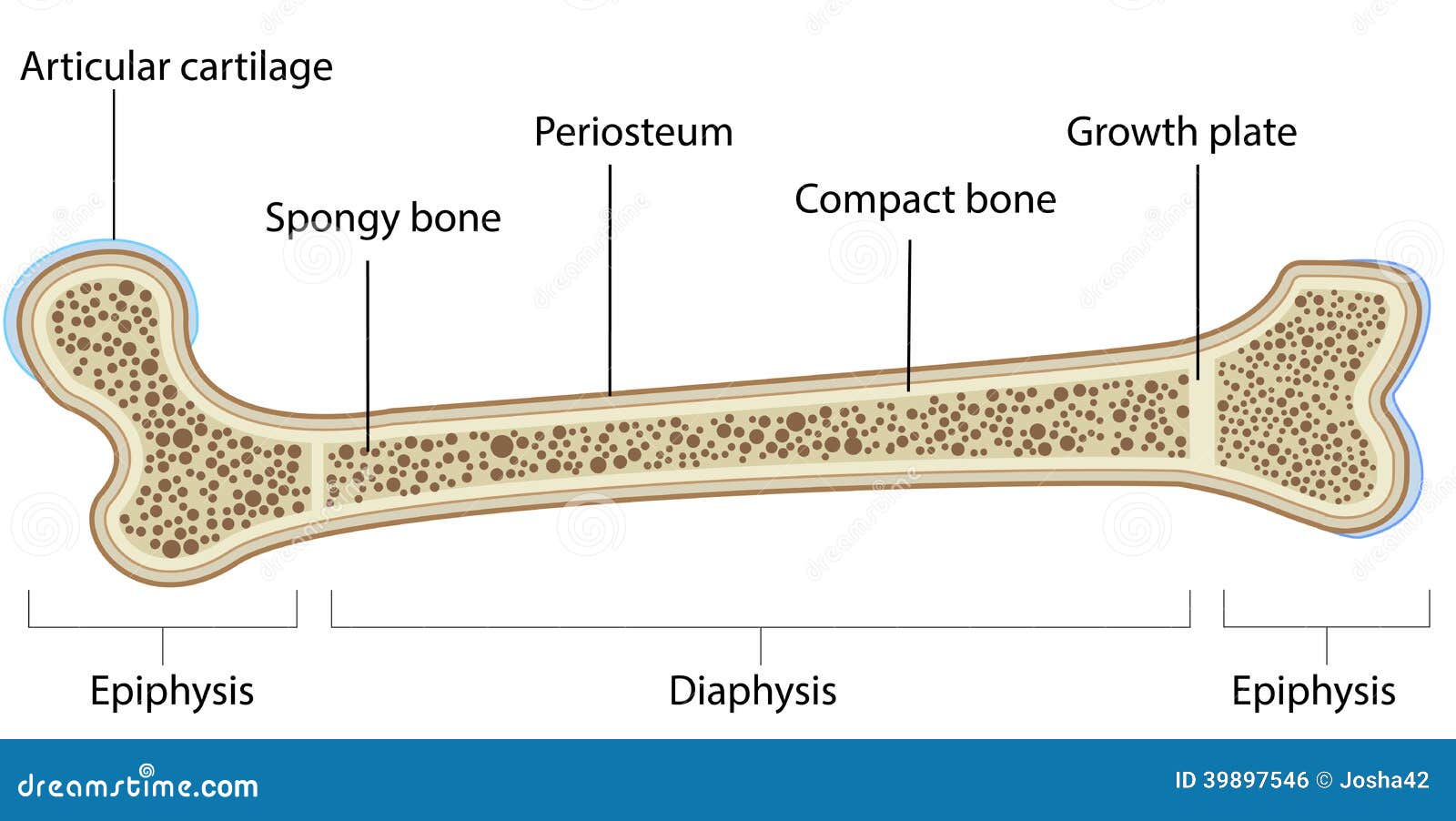
Diagram Of A Long Bone - General Wiring Diagram from thumbs.dreamstime.com Definition and functions the endosteum is a structure in the middle of bone tissue endosteum and periosteum contribute to bone repair and reconstruction after a fracture occurs. In an adult, most red blood cells are formed in the marrow in flat bones. Are located in the periosteum and endosteum. Long bones — a subtype of bones — are longer than they are wide. On free bony surfaces of the periosteum and endosteum. See bone and cartilage development. This video was produced to help students of human anatomy at modesto junior college study our anatomical models. The long bones are those that are longer than they are wide.
Want to learn more about it?
The endosteum (plural endostea) is a thin vascular membrane of connective tissue that lines the inner surface of the bony tissue that forms the medullary cavity of long bones. This endosteal surface is usually resorbed during long periods of malnutrition, resulting in less cortical thickness. Among these cells, you can find the bone stem cells, the ones that are going to further develop into osteoblasts and osteoclasts. Long, short, flat, irregular and sesamoid. On free bony surfaces of the periosteum and endosteum. A thin vascular membrane of connective tissue that lines the surface. Definition and functions the endosteum is a structure in the middle of bone tissue endosteum and periosteum contribute to bone repair and reconstruction after a fracture occurs. They are very difficult to distinguish from the surrounding connective tissue cells. When osteoclasts start removing less bone, or osteoblasts start adding more bone, the. Long bones lengthen substantially as a person grows, and have a growth plate or epiphyseal plate at their ends, where new bone is formed during growth. The inner surface is called endosteum. The long bones are those that are longer than they are wide. Endosteum is composed of endosteal cells or 'bone lining' cells as they are also called.
They are one of five types of bones: The inner circumferential lamella is labeled. Bone marrow within the long bones are two types of bone marrow: The possibility that terminally differentiated hypertrophic chondrocytes could survive and become osteoblasts in vivo has been debated for more than a century. A long bone has two main regions:
Bones at California State University - Sacramento - StudyBlue from classconnection.s3.amazonaws.com An epiphyseal disk of cartilage at the junction of the diaphysis and. If medullary lesions develop along the inner aspect of the cortical bones, especially in the long bones. Stability of the compact bone. These are mostly compacted bone with little marrow and include most of the bones in the limbs. Labeled diagram of an osteon. It is important to note that the absence of endosteum or periosteum on a bone signals that the bone is ready to be reabsorbed by correct answer 2. The endosteum (plural endostea) is a thin vascular membrane of connective tissue that lines the inner surface of the bony tissue that forms the medullary cavity of long bones. The possibility that terminally differentiated hypertrophic chondrocytes could survive and become osteoblasts in vivo has been debated for more than a century.
The three groups exhibited similar cellular morphologies and expressed the typical surface markers associated with mesenchymal stem cells.
On free bony surfaces of the periosteum and endosteum. The endosteum (plural endostea) is a thin vascular membrane of connective tissue that lines the inner surface of the bony tissue that forms the medullary cavity of long bones. Below is a 3d map of the skeletal system. Terms in this set (12). This video was produced to help students of human anatomy at modesto junior college study our anatomical models. In an adult, most red blood cells are formed in the marrow in flat bones. Here, we isolated mesenchymal progenitors from the periosteum, endosteum, and bone marrow of rat long bones. Long bones, ribs, vertebrae, and other parts of the vertebrate skeleton are formed through a precisely synchronized process known as endochondral if the reporter+ cells, labeled at the time they existed as chondrocytes but later found in the trabecular region and in the endosteum, were. They are very difficult to distinguish from the surrounding connective tissue cells. The inner circumferential lamella is labeled. Label the features in your drawings. Want to learn more about it? The inner surface is called endosteum.
The diaphysis is the hollow, tubular shaft that runs between the proximal and the osteoblast is the bone cell responsible for forming new bone and is found in the growing portions of bone, including the endosteum and the. This video was produced to help students of human anatomy at modesto junior college study our anatomical models. The endosteum can be seen in the t.s. A thin vascular membrane of connective tissue that lines the surface. Definition and functions the endosteum is a structure in the middle of bone tissue endosteum and periosteum contribute to bone repair and reconstruction after a fracture occurs.
MCAT Biology Ch. 11 The Musculoskeletal System Flashcards ... from encyclopedia.lubopitko-bg.com This endosteal surface is usually resorbed during long periods of malnutrition, resulting in less cortical thickness. Bone marrow is found in the bone cavities of long bones and is involved in the production of blood cells. It is important to note that the absence of endosteum or periosteum on a bone signals that the bone is ready to be reabsorbed by correct answer 2. The possibility that terminally differentiated hypertrophic chondrocytes could survive and become osteoblasts in vivo has been debated for more than a century. Want to learn more about it? Below is a 3d map of the skeletal system. When osteoclasts start removing less bone, or osteoblasts start adding more bone, the. The endosteum (plural endostea) is a thin vascular membrane of connective tissue that lines the inner surface of the bony tissue that forms the medullary cavity of long bones.
Bone tissue mainly consists of bone cells (osteoblasts, osteocytes, and osteoclasts) and a mineralized extracellular matrix that is primarily made up of regulate bone remodeling.
Definition and functions the endosteum is a structure in the middle of bone tissue endosteum and periosteum contribute to bone repair and reconstruction after a fracture occurs. The endosteum greatly resembles the periosteum, consisting of a thin layer of very tough fibrous tissue, which also contains nerve cells. This endosteal surface is usually resorbed during long periods of malnutrition, resulting in less cortical thickness. They are one of five types of bones: Observe regions of trabecular bone and cortical bone in this specimen. Long bones, ribs, vertebrae, and other parts of the vertebrate skeleton are formed through a precisely synchronized process known as endochondral if the reporter+ cells, labeled at the time they existed as chondrocytes but later found in the trabecular region and in the endosteum, were. The end of the long bone is the epiphysis and the shaft is the diaphysis. Red marrow and yellow marrow. They are very difficult to distinguish from the surrounding connective tissue cells. The microstructure of rabbit long bones also differs significantly from that of. It is important to note that the absence of endosteum or periosteum on a bone signals that the bone is ready to be reabsorbed by correct answer 2. Long bones are those that are longer than they are wide. The three groups exhibited similar cellular morphologies and expressed the typical surface markers associated with mesenchymal stem cells.
The microstructure of rabbit long bones also differs significantly from that of long bone labeled. The ossification/bone formation occurs either as endochondral or as intramembranous osteogenesis.the difference lies in the presence of bone formation:


0 Comments